Home

Contribution Assortment Inside fetal mca doppler loss curse Aggregate

Doppler indices of the umbilical and fetal middle cerebral artery at 18–40  weeks of normal gestation: A pilot study - ScienceDirect
Doppler indices of the umbilical and fetal middle cerebral artery at 18–40 weeks of normal gestation: A pilot study - ScienceDirect

Persistent reversed end‐diastolic flow of the middle cerebral artery: A  rare and concerning finding - Chainarong - 2022 - Journal of Clinical  Ultrasound - Wiley Online Library
Persistent reversed end‐diastolic flow of the middle cerebral artery: A rare and concerning finding - Chainarong - 2022 - Journal of Clinical Ultrasound - Wiley Online Library

A study of different scenarios of fetal middle cerebral artery peak  systolic velocity in an Indian population
A study of different scenarios of fetal middle cerebral artery peak systolic velocity in an Indian population

Middle Cerebral Artery Doppler Ultrasound Interpretation / Ultrasound in  Fetal Growth Assessment - YouTube
Middle Cerebral Artery Doppler Ultrasound Interpretation / Ultrasound in Fetal Growth Assessment - YouTube

OB Images
OB Images

US Pregnancy - Fetal Duplex - MCA Waveform - normally the MCA has a high  resistance flow - In chronic fetal h… | Cardiac sonography, Medical  ultrasound, Ultrasound
US Pregnancy - Fetal Duplex - MCA Waveform - normally the MCA has a high resistance flow - In chronic fetal h… | Cardiac sonography, Medical ultrasound, Ultrasound
Role of Doppler Sonography in Obstetrics | Radiology Key

Peak Systolic Blood Flow in the MCA

Peak Systolic Blood Flow in the MCA

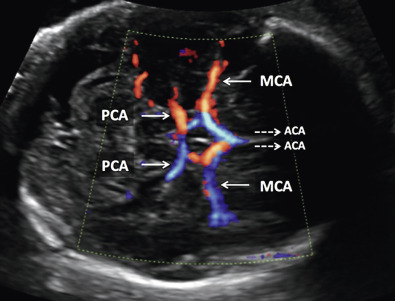
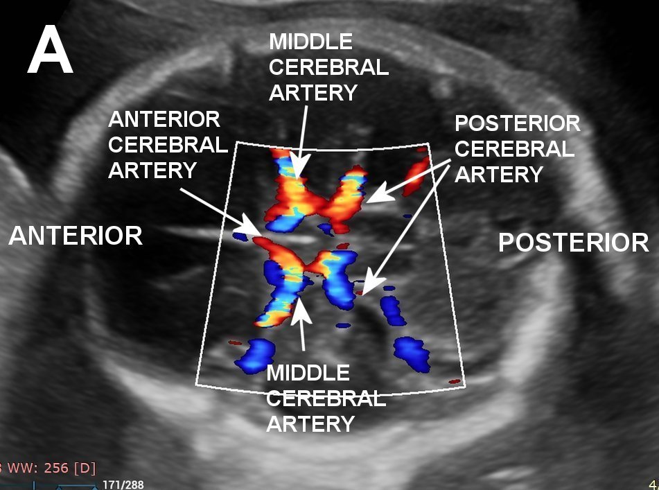
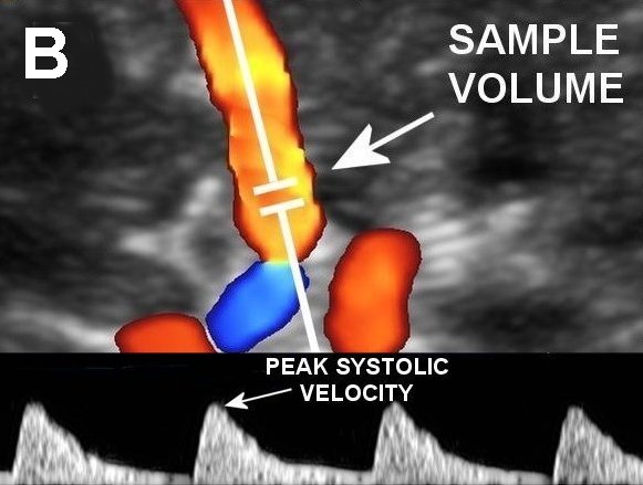
Fetal middle cerebral arterial Doppler assessment | Radiology Reference  Article | Radiopaedia.org
Fetal middle cerebral arterial Doppler assessment | Radiology Reference Article | Radiopaedia.org

Middle Cerebral Artery Doppler - ppt video online download

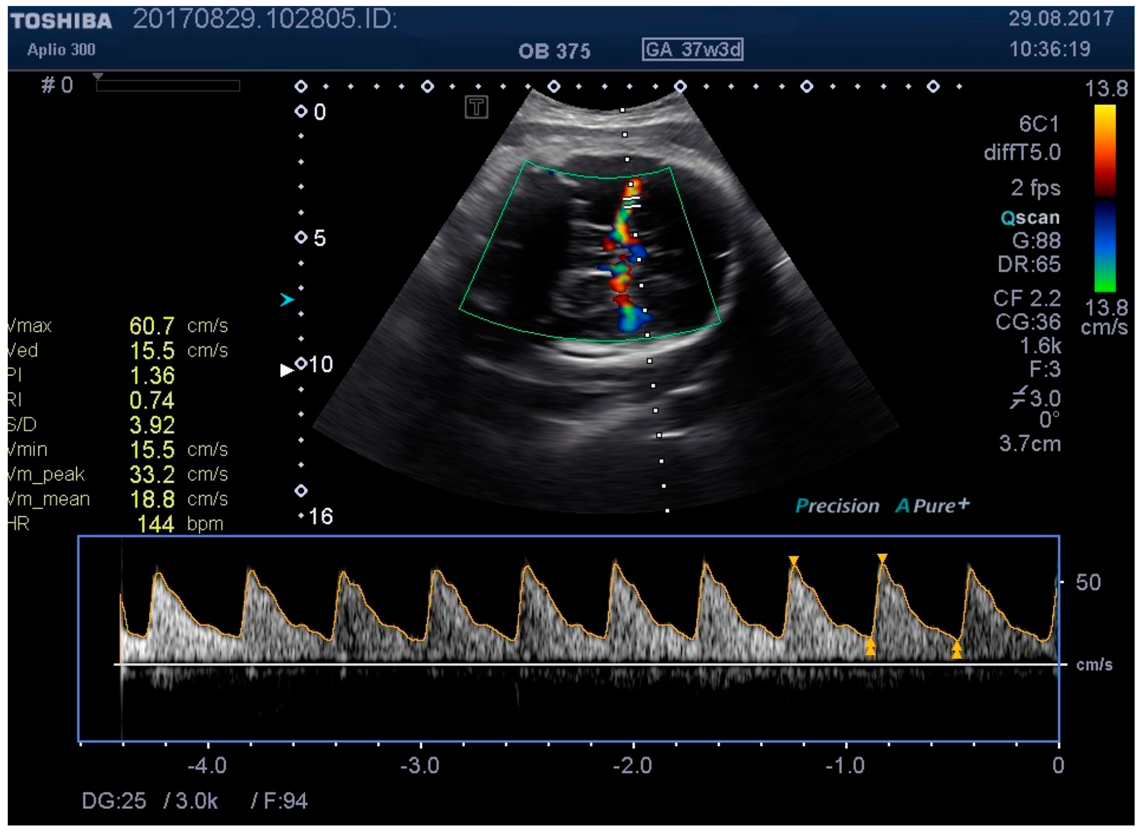
Medicina | Free Full-Text | Ultrasound Probe Pressure on the Maternal  Abdominal Wall and the Effect on Fetal Middle Cerebral Artery Doppler  Indices
Medicina | Free Full-Text | Ultrasound Probe Pressure on the Maternal Abdominal Wall and the Effect on Fetal Middle Cerebral Artery Doppler Indices
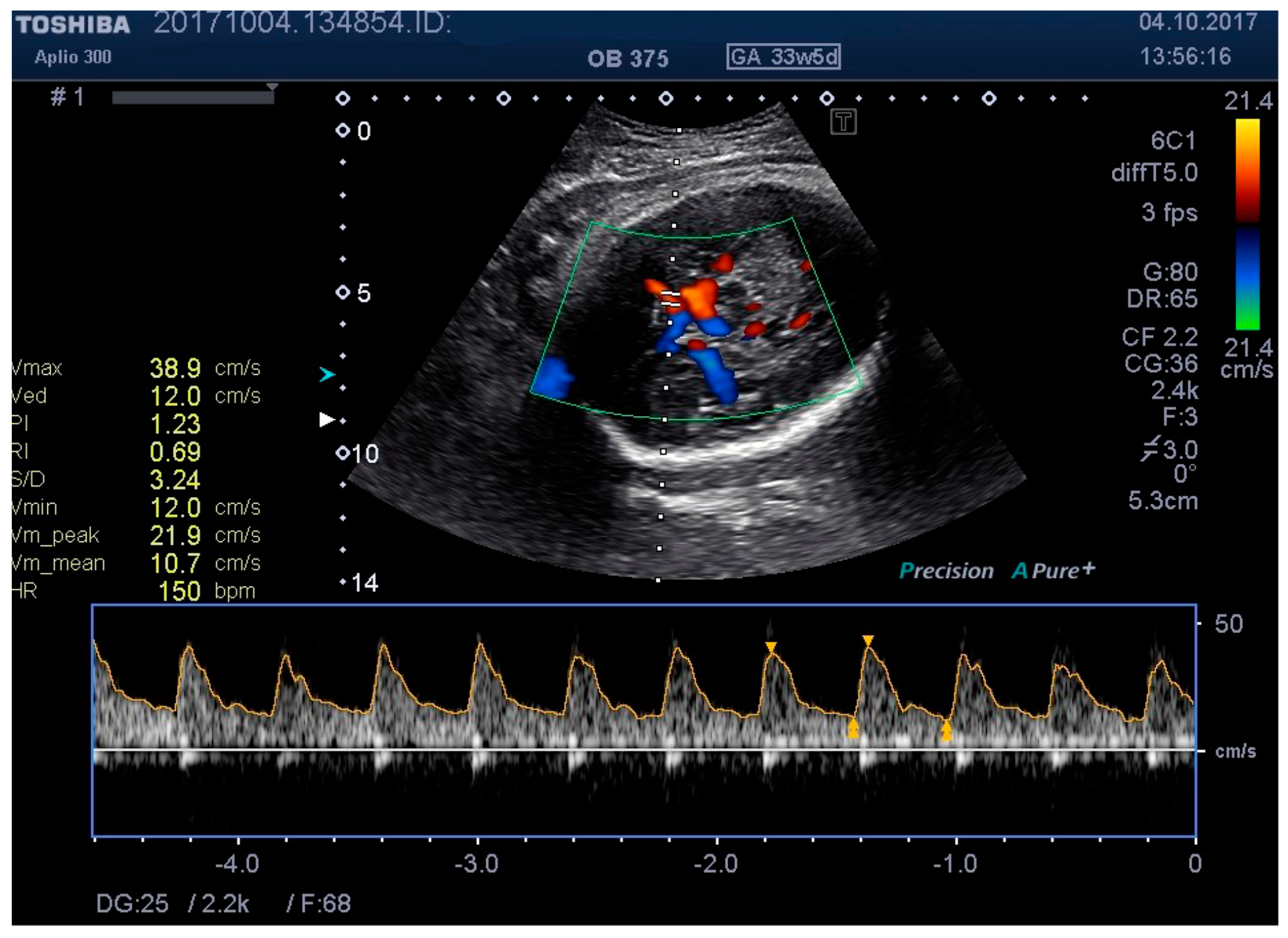
Medicina | Free Full-Text | Ultrasound Probe Pressure on the Maternal  Abdominal Wall and the Effect on Fetal Middle Cerebral Artery Doppler  Indices
Medicina | Free Full-Text | Ultrasound Probe Pressure on the Maternal Abdominal Wall and the Effect on Fetal Middle Cerebral Artery Doppler Indices
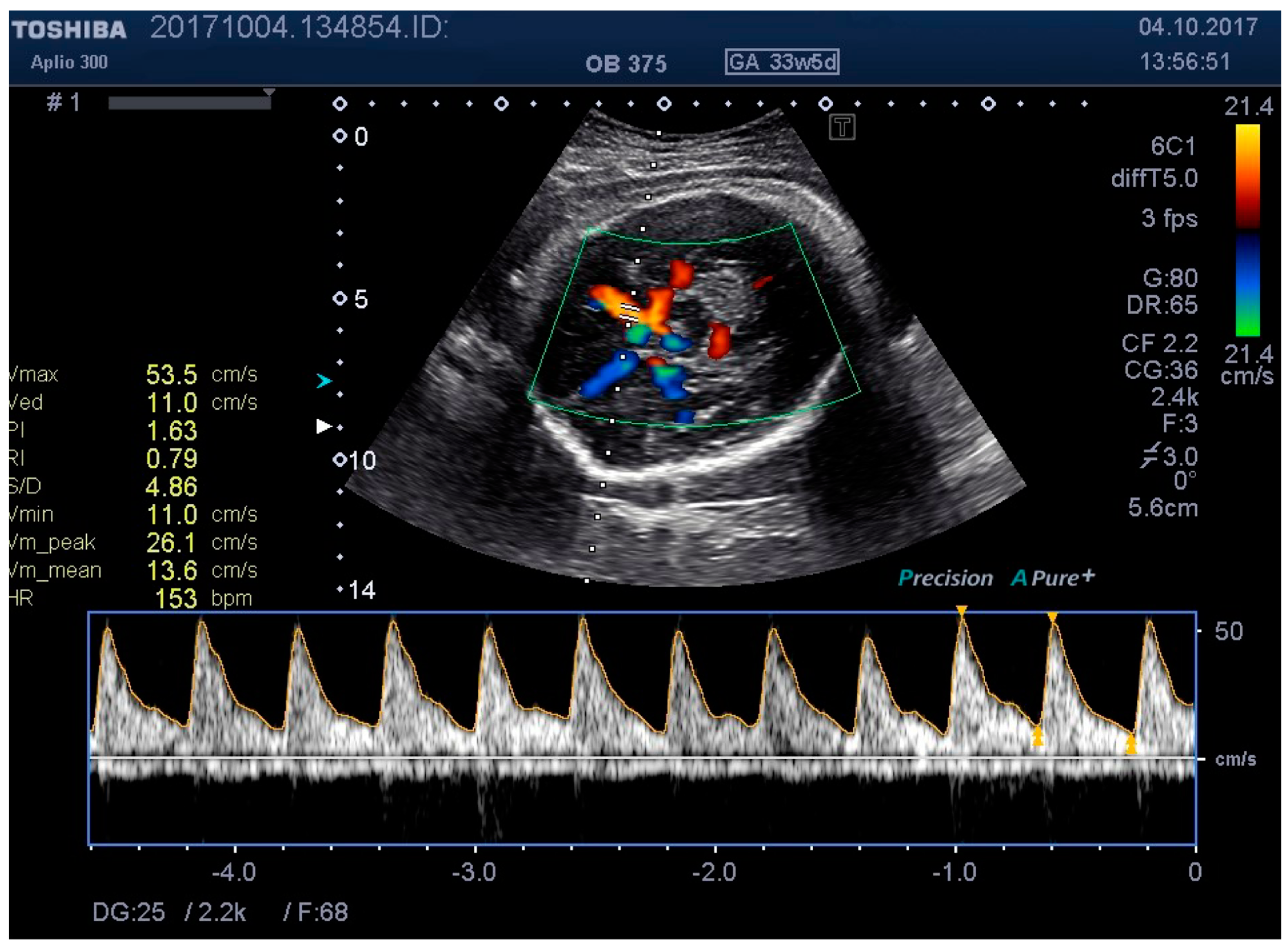
Medicina | Free Full-Text | Ultrasound Probe Pressure on the Maternal  Abdominal Wall and the Effect on Fetal Middle Cerebral Artery Doppler  Indices
Medicina | Free Full-Text | Ultrasound Probe Pressure on the Maternal Abdominal Wall and the Effect on Fetal Middle Cerebral Artery Doppler Indices

A classification of patterns of fetal middle cerebral artery velocity  waveforms as seen on Doppler ultrasound | Japanese Journal of Radiology
A classification of patterns of fetal middle cerebral artery velocity waveforms as seen on Doppler ultrasound | Japanese Journal of Radiology

Utility of Doppler parameters at 36-42 weeks' gestation in the prediction  of adverse perinatal outcomes in appropriate-for-gestational-age fetuses. -  Document - Gale Academic OneFile
Utility of Doppler parameters at 36-42 weeks' gestation in the prediction of adverse perinatal outcomes in appropriate-for-gestational-age fetuses. - Document - Gale Academic OneFile

Figure 9 from Applications of Doppler ultrasound during labor. | Semantic  Scholar
Figure 9 from Applications of Doppler ultrasound during labor. | Semantic Scholar

Fetal middle cerebral artery Doppler analysis | Download Table

Umbilical and Middle Cerebral Artery Doppler Measurements in Fetuses With  Congenital Heart Block - ScienceDirect
Umbilical and Middle Cerebral Artery Doppler Measurements in Fetuses With Congenital Heart Block - ScienceDirect

Example of fetal middle cerebral artery (MCA) Doppler with a good image...  | Download Scientific Diagram
Example of fetal middle cerebral artery (MCA) Doppler with a good image... | Download Scientific Diagram

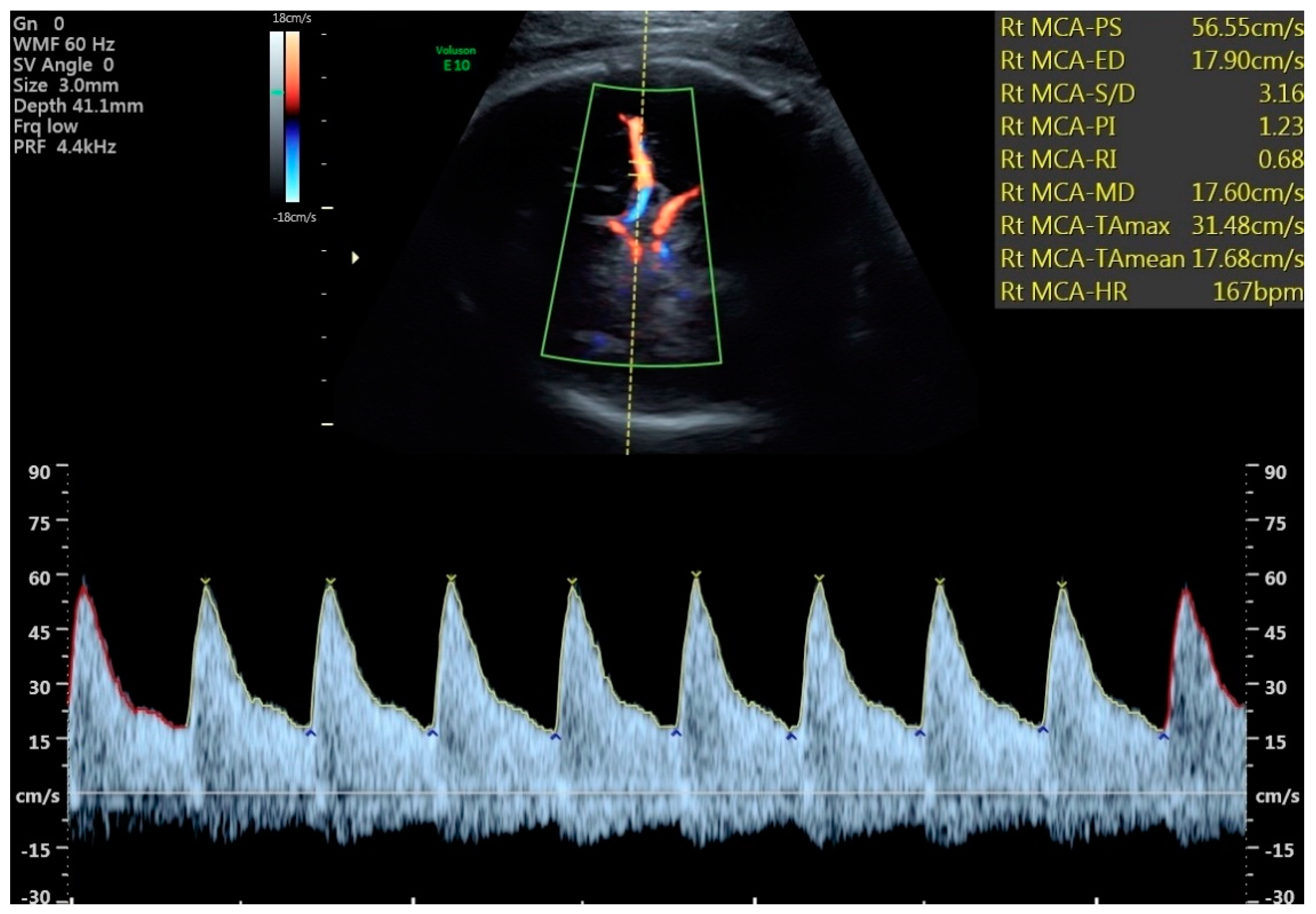
Blood flow spectra of fetal MCA. (a) Doppler waveform of MCA blood flow...  | Download Scientific Diagram
Blood flow spectra of fetal MCA. (a) Doppler waveform of MCA blood flow... | Download Scientific Diagram

Obstetric Doppler Part 2 - YouTube

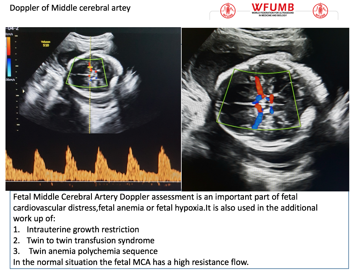
Ultrasound the Best #10: Doppler of the Fetal Middle of the Cerebral Artery  – WFUMB
Ultrasound the Best #10: Doppler of the Fetal Middle of the Cerebral Artery – WFUMB

A classification of patterns of fetal middle cerebral artery velocity  waveforms as seen on Doppler ultrasound | Japanese Journal of Radiology
A classification of patterns of fetal middle cerebral artery velocity waveforms as seen on Doppler ultrasound | Japanese Journal of Radiology
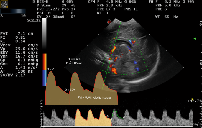
JCM | Free Full-Text | Patterns of Brain Sparing in a Fetal Growth  Restriction Cohort
JCM | Free Full-Text | Patterns of Brain Sparing in a Fetal Growth Restriction Cohort

Diagnostic and predictive value of Doppler ultrasound for evaluation of the  brain circulation in preterm infants: a systematic review | Pediatric  Research
Diagnostic and predictive value of Doppler ultrasound for evaluation of the brain circulation in preterm infants: a systematic review | Pediatric Research

Elevated fetal middle cerebral artery peak systolic velocity in diabetes  type 1 patient: a case report
Elevated fetal middle cerebral artery peak systolic velocity in diabetes type 1 patient: a case report

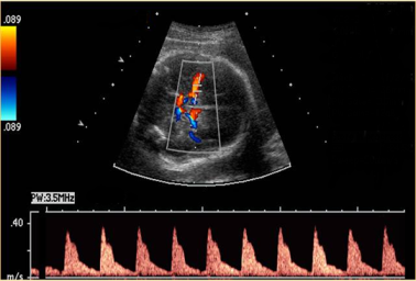
Ultrasound angiology reference standards of fetal cerebroplacental flow in  normal Egyptian gestation: statistical analysis of one thousand  observations | Egyptian Journal of Radiology and Nuclear Medicine | Full  Text
Ultrasound angiology reference standards of fetal cerebroplacental flow in normal Egyptian gestation: statistical analysis of one thousand observations | Egyptian Journal of Radiology and Nuclear Medicine | Full Text